| The eyeball is located in a bony socket called the orbit,
where it is suspended and surrounded by fat and blood vessels
together with motor and sensory nerves, including the optic nerve.
Six small muscles are attached to each eye enabling coordinated
movement of the pair. The adult human eye is approximately 2.5 cm in
diameter. Definitions:
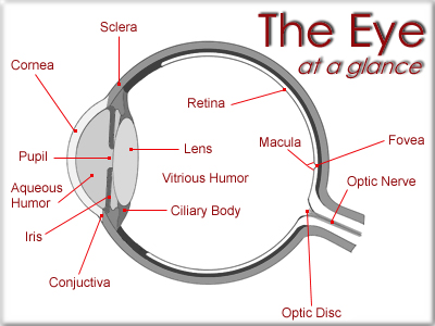
• The Sclera - The white outer part of the eye. It is a tough
external coat. Six muscles attached to the sclera enable the eye to
look up, down and side-to-side. Part of the sclera forms a clear
window, the cornea, through which light gets into the eye.
• The Cornea - The clear outer part of the eye's focusing system
located at the front of the eye. Most of the bending of the light
rays (refraction) occurs at the cornea. The lens also bends the
light but to a lesser extent. The lens does a sort of fine tuning to
insure that the image is sharply focused on the retina.
• Aqueous Humor - The clear watery fluid filling the anterior
chamber of the eye between the cornea and the lens. It has the
benefit of being fairly homogenous and, the optical properties are
easily measured. The space that it inhabits is called the anterior
chamber.
• Vitreous Humor - The clear gel filling the posterior chamber of
the eye between the lens and the retina. The space that it fills is
called the vitreous body.
• The Pupil - The opening at the center of the iris. The iris
adjusts the size of the pupil and controls the amount of light that
can enter the eye.
• The Iris - The colored tissue behind the cornea - color varies
from pale blue to dark brown.
• The Choroid - A spongy layer filled with blood vessels. It lies
between the sclera and the retina. The choroid nourishes the outer
layers of the retina.
• The Conjunctiva - The transparent mucous membrane lining the
inside of the eyelids and the white of the eyeball.
• The Macula - The small sensitive area of the retina that gives
central vision; contains the fovea. This foveal area is covered with
a yellow pigment called the macula lutea.
• The Optic Nerve - The bundle of over one million nerve fibers that
carries visual messages from the retina to the brain.
• The Fovea - The center of the macula; gives the sharpest vision.
When we fixate or look directly at an object it is imaged on the
fovea.
• The Lens - The lens is the clear part of the eye behind the iris
that helps to focus light on the retina. The lens helps to focus on
both far and near objects so that they are perceived clearly and
sharply. The ciliary muscle helps to change the shape of the lens.
This changing of lens shape is called accommodation.
The lens itself is a multilayered structure (something like an
onion). In young people it is normally perfectly clear and quite
elastic. As one ages its elasticity is reduced. In fact after the
age of about 45 the lens' ability to change in shape is considerably
reduced. That is why people over the age of 45 almost always require
glasses to read and/or to see distant objects. It is not unusual for
people in their 50's and older to wear bi-focal or even tri-focal
lenses.
As one gets older the lens can also become cloudy. This condition is
called cataract. When cataracts become too severe the lens has to be
removed and be replaced with an artificial lens. Of course the
artificial lens is not capable of accommodation. However, by the
time most people are afflicted with cataracts they will already be
old enough so that they will have naturally lost most of their
accommodation. Also as one ages the lens becomes more yellow. People
in their 50s will, for example, exhibit clearly lower spectral
sensitivities at the short wavelength end of the spectrum than 10
year olds.
• The Ciliary Body - The Ciliary Body consists of bundles of tiny
muscles used in accommodation. When the ciliary muscle is relaxed,
the choroid acts like a spring pulling on the lens via the zonule
fibers causing the lens to become flat. When the ciliary muscle
contracts, it stretches the choroid, releasing the tension on the
lens and the lens becomes thicker.
• The Retina - The light-sensitive tissue lining the back of the
eyeball; sends electrical impulses to the brain. On the right is a
photo of what an eye-care person sees when looking at your retina
with an ophthalmoscope. The dark area near the center is the fovea .
This area is actually a depression in the retina. Although this
photo does not show it, the foveal area has a yellow pigmentation
called the macula lutea. When we fixate (look directly at) objects,
images of these objects are projected on to the fovea. It is the
retinal location of our best visual acuity and color vision.
The the optic disc is the place where all the blood vessels and
optic nerves converge and go out of the retina to the brain. The
optic disc, also called the blind spot, is where the axons of the
ganglion cells leave the retina to form the optic nerve.
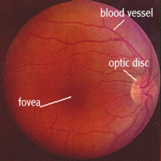
It is called the blind spot because there are no rod or cone
receptors in this part of the retina and we can not see objects that
are imaged on this part of the retina.
Summation:
Sight is our most precious sense, and any diminution of it is truly
regrettable. Our eyes alone save us from perpetual darkness, and
they fill our lives with all the glory of creation. They enable us
to scan the vast sweep of the universe a million light years away,
or examine the minute details of a flower held in the hand.
Our eyes are wondrous devices that we often take for granted, too
often fail to protect, and rarely understand.
It is important that you develop a good relationship with your eye
care practitioner, because you may be seeing a great deal of him or
her over a long period of time. You should have confidence in
him/her and find it comfortable to ask questions.
Eye care practitioners are often very busy, but they should never be
too busy to discuss your case with you. Find an eye care
practitioner with whom you are comfortable then stay with him/her.
Your eye care practitioner needs your cooperation. He/she has no
control over what happens between office visits, you have. Think of
him/her as a partner in the care of your eyes, but remember that you
are primarily responsible for your own welfare. He/she is a resource
person, a consultant who will prescribe a program of treatment and
/or the medications to be used in that program.
Implementing the program is up to you. If you do not care diligently
for your eyes it is unrealistic to expect anyone else to do so.
Surgery is normally suggested only if you cannot see well enough
with your contact lenses to drive or function in your work, or if
you are unable to wear contact lenses. If you are not sure about
surgery when it is recommended, seek a second, independent opinion.
If your eye care practitioner advises you to see him/her again at a
certain time, keep the appointment. There is a good reason for it,
and if you wonder what it is, ask them.
Inform any physicians that you may be seeing about any eye problem
and any medication you are taking. Be alert to any changes in your
eye condition or in your vision. If you experience blurring,
scratchiness, irritation, watering or any discharge, you must
contact your eye care practitioner immediately. This may signal a
problem with your eyes' tolerance of your contact lenses or the need
for refitting.
You should, or course, take normal care of your eyes and avoid the
use of any substance that is not prescribed by your eye care
practitioner.
|